Peptides can be tagged with various fluorescent dyes at various positions. Fluorescent labeling of peptides can be used for microscopy, FACS, in-vivo fluorescent imaging…
Available dyes
SB-PEPTIDE offers a wide range of fluorescent labeling options to answer your needs.
FAM and FITC are fluorescein derivatives. FAM is a carboxyfluorescein which shows an absorption at 495nm and an emission at 517 nm. FAM is the most commonly used fluorescent dye attached to peptides. Moreover, FAM is used with most fluorescence detection equipment. FAM is, most often, used in the pH of 7.5 to 8.5 and can be attached to amino or carboxy-terminal. This fluorescein is used in the sequencing of nucleic acids and in the labeling of nucleotides.
TAMRA: tetramethylrhodamine is a rhodamine derivative. TAMRA is most commonly used for preparing bioconjugates, such as fluorescent antibody used in immunochemistry studies and cellular imaging. But TAMRA is also used for oligonucleotide labeling and DNA sequencing. TAMRA have an excitation peak at 552nm and an emission peak at 578nm.
Cyanine dyes are used in biotechnology. Indeed, cyanine dyes are famous in genomic hybridization, in transcriptomics, in proteomics studies like FRET, immunoassays… Cyanine dyes are used to label peptides, proteins, antibodies for fluorescence detection techniques such as flow cytometry, microscopy, microarrays… Cyanine dyes can have different modifications which confer different excitation and emission wavelength. The most commonly used is Cy3 and Cy5 with respectively wavelength: excitation at 550nm, emission 570nm, and excitation at 650nm and emission at 670nm.
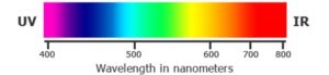
ATTO dyes are also used in biotechnology. Indeed, ATTO dyes are most used in labeling DNA, RNA, proteins, peptides in particular for fluorescence microscopy. ATTO dyes have different excitation and emission wavelength. They cover a wide spectral range from 390nm (UV) up to 730nm (near infrared). This specificity allows to used most commonly light sources.
Alexa dyes can be used to label primary antibodies, secondary antibodies to amplify signal and sensitivity. Alexa dyes can also be conjugated to proteins and peptides. Like ATTO dyes, Alex dyes covers a spectral emission and excitation range in from visible spectrum up to the infrared spectrum.
SiR is a fluorogenic dye which is cell permeable and highly specific live cell DNA probe. SiR dye is most used for fluorescence imaging of the nucleus and DNA and can be conjugated to peptides.
STAR635 is a fluorescent dye which can be conjugated to peptides. STAR635 is commonly used for STED application. This dye have an excitation peak at 635nm and an emission peak at 655nm.
Various positions can be labeled: N-ter, side chain of specific lysines…
Fluorescent dyes for cell penetration monitoring
Some fluorescent dyes are conjugated to a variety of antibodies, peptides and proteins optimized for cellular labeling and detection. Such as:
Alexa 488 dye (excitation at 490nm, emission at 525nm)
Alexa 555 dye (excitation at 555nm, emission at 580nm
Tetramethylrhodamine (TRITC) dye (excitation at 557nm, emission at 576 nm)
SiR dye (excitation 650nm, emission 670nm)
Cell-penetration peptide assessment
The fluorescent labeling of peptides is also used to detect the cell penetration of a given peptide by microscopy. SB-PEPTIDE offers to conjugate your peptides to a fluorescent CPP of your choice among TAT, Maurocalcine, penetratin...
FRET peptides
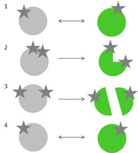
FRET peptides are used to study the binding of peptides to proteins (1), conformational change of peptides (2), protease activity (3), measurement of the molecular proximity (4).
FRET fluorophore/quencher pairs examples:
Abz / Dnp (λexc=320nm, λem=420nm)
EDANS / Dabcyl (λexc= 340nm, λem= 490nm)
Mca / Dnp (λexc= 325nm, λem= 492n)
Trp / Dnp (λexc= 280nm, λem= 360nm)
FAM / Dabcyl (λex= 492nm, λem= 517nm)
TAMRA / BHQ3 (λex= 543 nm, λem= 572 nm)
Methods of conjugation:
Click chemistry: Fluorescent dyes can be conjugate to peptide by click chemistry. Click chemistry consist in of clipping two molecules to each other. The reaction involves an alkyne and a nitrogen-bearing functional group (azide). The peptide is modified to have one of these functions and to allow the reaction. The fluorescent dye used have the second function. The reaction is catalyzed with a molecule of cupric. Click chemistry is a conjugation method which is very selective, effective, quick, quantitative, takes place in water and easy to set up.
IMAGE A: fluorescent dye-click chemistry
Image B: Click chemistry provides an efficient and mild way to make triazole‑stapled peptides through on‑peptide macrocyclization. By introducing azide‑ and alkyne‑containing unnatural amino acids such as L‑Nle(ε‑N₃) and D‑propargylalanine (D‑Pra), a single triazole staple can be formed via copper‑catalyzed azide–alkyne cycloaddition, giving conformationally constrained peptides with improved properties.
Amide bond: Fluorescent dyes can be conjugate to peptide by amide bond using carboxylated or NHS ester functionalized dye which will bond to free amines. This reaction implies an amino moiety to bind the peptide with the dye. Thanks to selective protective groups, SB-PEPTIDE can label specifically one free amine in case a peptide contains several free amines.
Maleimide-thiol: conjugation on free thiol…
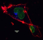
Localization studies
A peptide that targets another protein can be fluorescently labeled in order to detect the expression of the ligand or to localize/co-localization it in a tissue by confocal microscopy for instance.
Fluorescent peptides for FACS
Fluorescent peptides can also be useful as fluorescent probes to measure the proliferation of cell population in FACS which express a ligand of interest.
Biodistribution studies
Peptides can be fluorescently tagged to evaluate their biodistribution in-vivo during the pre-clinical development.
1- Birch D et al. Biochim Biophys Acta Biomembr. 1859(12):2483-2494 (2017)
Cell-penetrating peptides constitute efficient delivery vectors, and studies of their uptake and mechanism of translocation typically involve fluorophore-labeled conjugates. In the present study, the influence of a number of specific fluorophores on the physico-chemical properties and uptake-related characteristics of penetratin were studied. An array of seven fluorophores belonging to distinct structural classes was examined, and the impact of fluorophore labeling on intracellular distribution and cytotoxicity was correlated to the physico-chemical properties of the conjugates. Exposure of several mammalian cell types to fluorophore-penetratin conjugates revealed a strong structure-dependent reduction in viability (1.5- to 20-fold lower IC50 values as compared to those of non-labeled penetratin). Also, the degree of less severe effects on membrane integrity, as well as intracellular distribution patterns differed among the conjugates. Overall, neutral hydrophobic fluorophores or negatively charged fluorophores conferred less cytotoxicity as compared to the effect exerted by positively charged, hydrophobic fluorophores. The latter conjugates, however, exhibited less membrane association and more clearly defined intracellular distribution patterns. Thus, selection of the appropriate flurophore is critical.
Fluorescence resonance energy transfer (FRET) detects the proximity of fluorescently labeled molecules over distances >100 A. When performed in a fluorescence microscope, FRET can be used to map protein-protein interactions in vivo. We here describe a FRET microscopy method that can be used to determine whether proteins that are colocalized at the level of light microscopy interact with one another. This method can be implemented using digital microscopy systems such as a confocal microscope or a wide-field fluorescence microscope coupled to a charge-coupled device (CCD) camera. It is readily applied to samples prepared with standard immunofluorescence techniques using antibodies labeled with fluorescent dyes that act as a donor and acceptor pair for FRET. Energy transfer efficiencies are quantified based on the release of quenching of donor fluorescence due to FRET, measured by comparing the intensity of donor fluorescence before and after complete photobleaching of the acceptor. As described, this method uses Cy3 and Cy5 as the donor and acceptor fluorophores, but can be adapted for other FRET pairs including cyan fluorescent protein and yellow fluorescent protein.
3- Medintz I L et al. Nat Mater. 5(7):581-9 (2006)
Proteases are enzymes that catalyse the breaking of specific peptide bonds in proteins and polypeptides. They are heavily involved in many normal biological processes as well as in diseases, including cancer, stroke and infection. In fact, proteolytic activity is sometimes used as a marker for some cancer types. Here we present luminescent quantum dot (QD) bioconjugates designed to detect proteolytic activity by fluorescence resonance energy transfer. To achieve this, we developed a modular peptide structure which allowed us to attach dye-labelled substrates for the proteases caspase-1, thrombin, collagenase and chymotrypsin to the QD surface. The fluorescence resonance energy transfer efficiency within these nanoassemblies is easily controlled, and proteolytic assays were carried out under both excess enzyme and excess substrate conditions. These assays provide quantitative data including enzymatic velocity, Michaelis-Menten kinetic parameters, and mechanisms of enzymatic inhibition. We also screened a number of inhibitory compounds against the QD-thrombin conjugate. This technology is not limited to sensing proteases, but may be amenable to monitoring other enzymatic modifications.
Cell-permeable peptides were evaluated for a quantitatively controlled import of small molecules. The dependence of the import efficiency on the fluorophore, on the position of the fluorophore as well as on the nature of the cargo were addressed. Cellular uptake was quantitated by flow cytometry and fluorescence correlation microscopy (FCM). Fluorophores with different spectral characteristics, covering the whole visible spectral range, were selected in order to enable the simultaneous detection of several cell-permeable peptide constructs. The transcytosis sequences were based either on the sequence of the Antennapedia homeodomain protein (AntpHD)-derived penetratin peptide or the Kaposi fibroblast growth factor (FGF)-derived membrane translocating sequence (MTS)-peptide. In general, the AntpHD-derived peptides had a three- to fourfold higher import efficiency than the MTS-derived peptides. In spite of the very different physicochemical characteristics of the fluorophores, the import efficiencies for analogues labelled at different positions within the sequence of the import peptides showed a strong positive correlation. However, even for peptide cargos of very similar size, pronounced differences in import efficiency were observed. The use of cell-permeable peptide/cargo constructs for intracellular analyses of structure-function relationships therefore requires the determination of the intracellular concentrations for each construct individually.
Cookies settings
This website uses cookies to ensure the operation and security of the website.
If you click on “Accept all”, we and third-party providers will process your personal data and store information (e.g. through cookies) on your device or access it. By clicking on the “Settings” button, you can view details of the processing, set your preferences, and reject processing operations that require consent.
Functional
Toujours activé
The technical storage or access is strictly necessary for the legitimate purpose of enabling the use of a specific service explicitly requested by the subscriber or user, or for the sole purpose of carrying out the transmission of a communication over an electronic communications network.
Préférences
The technical storage or access is necessary for the legitimate purpose of storing preferences that are not requested by the subscriber or user.
Statistiques
The technical storage or access that is used exclusively for statistical purposes.The technical storage or access that is used exclusively for anonymous statistical purposes. Without a subpoena, voluntary compliance on the part of your Internet Service Provider, or additional records from a third party, information stored or retrieved for this purpose alone cannot usually be used to identify you.
Marketing
The technical storage or access is required to create user profiles to send advertising, or to track the user on a website or across several websites for similar marketing purposes.